Posted on May 24th, 2017
What is cholesterol and where does its name come from? Wikipedia has the following to say about cholesterol: The name cholesterol comes from the Ancient Greek word chole-(bile) and stereos (solid), followed by the chemical suffix-ol, which is for alcohol. Cholesterol is a steroid or modified sterol (1), a type of lipid molecule that is […]
Posted on May 22nd, 2017
Interestingly enough, women that are on estrogen supplements often experience no significant thyroid changes, however about 40 % of these women that were also taking thyroid medication, did experience a reduction in their T4 (thyroxin) levels and did begin to show signs of hypothyroidism. The June 7, 2016 by the New England Journal Medicine found […]
Posted on May 20th, 2017
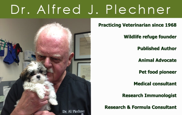
There is so much information known about treating the effects of autoimmunity and cancer in animals and in humans, and definite thoughts about why these diseases occur, but no real measurable cause. Over the past 50 years of practice, I have discovered an endocrine immune imbalance that seems to be the cause of autoimmunity and […]
Posted on May 18th, 2017
What is testosterone? Testosterone is a masculine hormone that is found both in men and in women. Several medical studies on the internet indicate that testosterone is usually produced in one area of the body, and travels to other areas of the body and to other organs. Where is testosterone produced? It is produced in […]
Posted on May 16th, 2017
What is human growth hormone? HGH is a hormone produced by the pituitary gland. It functions to cause the liver to release a second hormone referred to as Insulin-Like Growth Factor (IGF-1) and is responsible for helping children gain height, improve their muscle mass and help other organs and tissue increase production of more of […]
Posted on May 14th, 2017
What is the internet definition of hypothyroidism? Hypothyroidism = abnormally low activity of the thyroid gland resulting in the retardation and development of children and adults. However, medical science has discovered many more clinical signs and clinical symptoms for patients that are suffering from hypothyroidism which include canines, felines and humans. What are the causes […]
Posted on May 12th, 2017
The radio host for Take Charge of Your Health, Corinne Funari, was kind enough to ask Dr. Plechner to join her and Carol Peterson in doing an interview on her radio program last month. According to Corinne, she received some very positive feedback and if you would like to listen to her interview, you are […]
Posted on May 11th, 2017
OBESITY: WHY HAS DIETING AND EXERCISE NOT HELPED? Many Americans and their pets have been facing obesity problems for many years. Strict dieting with special nutritional programs and special exercise programs seem to be of little value for many people that are trying to lose excess weight. Many people have consulted their physician for themselves […]
Posted on April 23rd, 2017
That simple enzyme that can induce catastrophic diseases for patients is called aromatase. According to Wikipedia, aromatase is an adrenal enzyme. It is also called estrogen synthetase or estrogen synthate. Aromatase has the ability to transform androstenedion into estrone and testosterone into estradiol. Aromatase is therefore responsible for aromatization of androgens into estrogens. This enzyme […]